Are your calf muscles even fit enough to run?
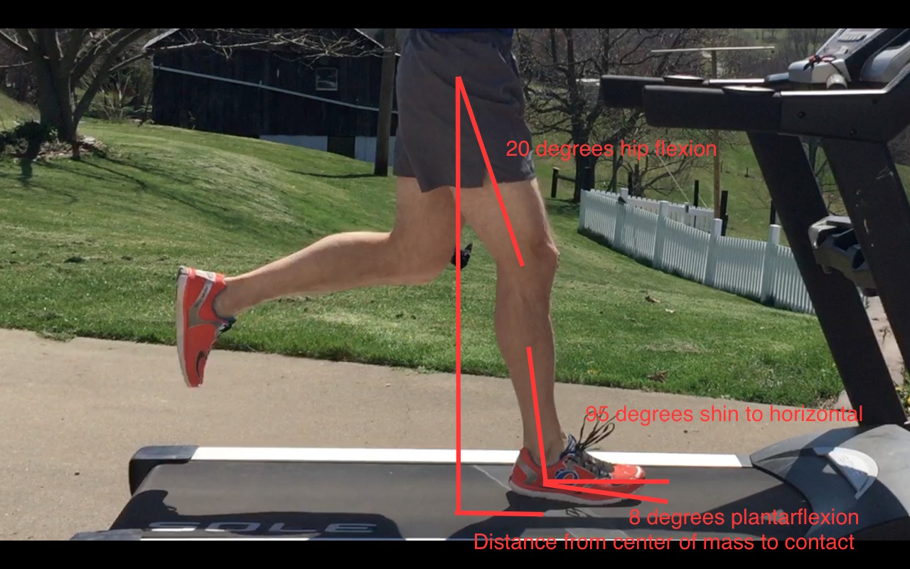
I’ve come to the conclusion that nearly everybody’s calf muscles stink. It’s partly a problem because runners tend to underestimate structural demand and think of the calf as a minor muscle group that doesn’t do much except push them a bit forward. Surely the big hip and thigh muscles are the real workers, right? Pushing is certainly some of the calf’s role during the later part of the running stride, in that brief moment before your foot leaves the ground. But slightly earlier in the stride, which is called mid-stance, as your body is moving forward over your fixed leg, the calf muscles are in full workhorse mode. 

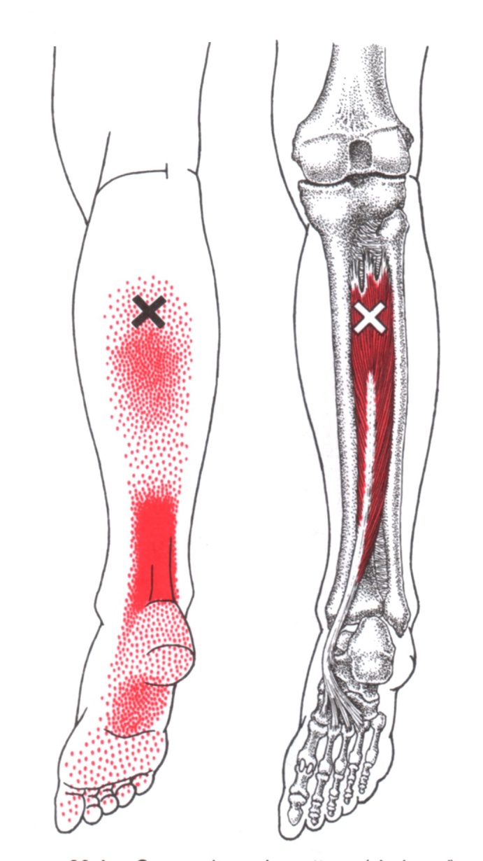
This mid-stance timing is actually the moment when the soleus, which is the biggest calf muscle, stealthily receives its peak demands. The soleus provides a major stabilizing effect to the ankle and knee, working synergistically with the thigh’s big quadriceps to keep gravity from crushing you down like Wile E. Coyote getting a boulder dropped on his head. The calf does this by resisting the forward momentum of your shin bone, using the foot as a point of fixation to work through. (Which is great if your foot does its job correctly but that’s a story for another day.) 

Stop worrying about the shoe marketing hype around pronation and cushioning and start worrying about getting stronger

In 2012, Dorn et al. calculated that the soleus produced an average peak of 6 times body weight force at a 7:40 min/mile pace (7.8 mph), a pace that’s arguably quicker than many recreational folks run as they trot along. By comparison, the quadriceps were producing a measly 4 times body weight in average peak force at that pace. Doesn’t seem fair that the big thigh muscles produce less force than the smaller calf muscles does it? In that same study, speeding up to 15.7 mph (3:49 min/mile pace), a full blown sprint to most of us, resulted in an average peak of more than 8 times body weight force demands in the soleus. I know what you are thinking. That you never have or never will run that fast. Me either. Because my calf muscles suck, just like yours. (Not true, I will beat you in any calf raise test that you challenge me to as long as it’s on the moon.) The real point is that your calf muscles work ridiculously hard, even at slower paces, and speeding up further elevates their demand. 

The researchers stated, “Across all running speeds, soleus, gastrocnemius and quadriceps provided roughly 75% of the total vertical support impulse needed to accelerate the body upward, with soleus contributing as much as 50%.” Notice the word vertical. The calf just wants to keep your leg tall, where your knee is bent just enough to absorb the blow of landing on the earth but not so bent that your quads work overtime or you linger on the ground too long. That’s why knee pain can relate back to less obvious factors like calf muscle function just as much, if not more so than the thigh muscles that are often blamed. And vice versa, Achilles tendon issues and calf strains can relate to poor thigh muscle function. 

Older runners, especially men over 35 who make up the masters crowd, are particularly impacted by calf and Achilles tendon injuries. One unfortunate issue with age is that the Achilles tendon reduces in stiffness. I know it sounds counterintuitive at first, but research, like that from Willy in 2019, indicates that older tendons tend to have increased compliance (stretchiness) while stiff tendons do a better job at transmitting force and are less likely to be overstretched in a way that mechanically overstrains the tissue. Would you rather drive across a suspension bridge supported by slightly forgiving but very stiff steel cables or very forgiving and less stiff rubber bands? In 2017 Mahieu et al. recognized a similar situation where weaker calf muscles were even associated with Achilles tendon injury in a group of young, male military officer cadets. 

Coupled with weakening calf muscles, an overzealous spring marathon training cycle can quickly turn to a puff of smoke. Once you’ve exceeded the structural capacity of the Achilles tendon and calf muscles, no amount of stretching is going to fix what you broke in that March moment of early season speedwork passion. As a side note, I really wish people would stop trying to stretch out tendon and muscle strain injuries. 

Even if a person hasn’t been injured and just laid low all winter to focus on slurping [insert your beer of choice], initially adding small amounts of medium to high speed strides is the safer option for a few weeks versus going out for 2 or more miles of speedwork. In order to keep the structural and neurological capacity at a level to tolerate the highest forces, I like to have athletes perform strides year round, even if they aren’t specifically doing periodized speedwork for a target event. Strides can be short, 5 to 10 second bouts before, during, or after aerobic runs but eventually can become much longer or performed uphill to encourage even more power production while reminding your tissues that they do have a very high stress job to perform. If an endurance athlete has been hurt for a while, getting consistency and volume back are the first priority with short, slow runs. Then we can start to titrate in a little speed with strides. None of these things are appropriate, though, if the calf muscle and Achilles tendon capacity are garbage after prolonged periods of missed runs. 

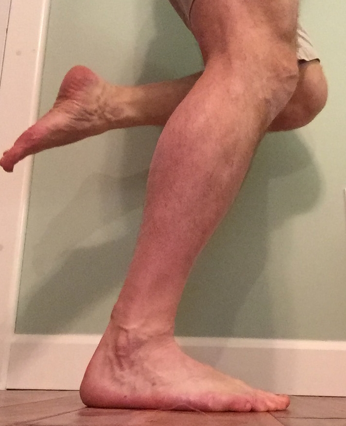
As for the nitty gritty of determining just how much your calf muscles stink? In 2017 Herbert-Losier et al. developed age-based normative values for the number of single leg calf raises that a person should be able to perform. These must be high quality (eg. full ankle motion with control) in order to count. You can see in the chart that the numbers are quite high. If I was still a spry young 30 year old, I should be able to complete 32 reps but now that I’m just about to hit that big 6-0 mark, my number is 23, which seems like a pretty high number to everyone whose last name isn’t Herbert-Losier. I’d estimate that fewer than half the people I see in the clinic are able to achieve their appropriate number. What’s your number? 

Data Adapted from Herbert-Losier et al. 2017

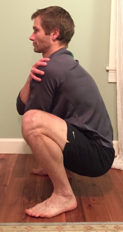
If you aren’t making the Herbert-Losier grade but aren’t currently injured, making it a point to isolate the calf for strengthening could pay off for injury prevention. There are no guarantees, because no one is immune to injury, but there is never a penalty for being stronger. For the complete beginner, strengthening doesn’t need to be complicated. A basic double leg calf raise, knees straight, from floor height could suffice for two to three times per week for two to three weeks. The goal is to feel fatigue in the calf muscle, not pain. Once that isn’t challenging, we progress to single leg straight knee calf raises and also start to do bent knee calf raises too. The soleus works hard in both positions so don’t lose any sleep about that. Once your muscles start to come out of their coma, which could still take a few more weeks, we work toward raises on steps and raises with extra weight to further increase the resistive and range of motion demands. 

Just for some perspective on how important calf strength generation is to me personally, I did single leg calf raises in straight and bent positions last week with 95# on my back for 15 repetitions in each position and of course on both legs. Sometimes I’ll do more weight, like 110-140# with reduced reps of 5-10 times. This isn’t where most runners can start and that could easily hurt many people so I’m not telling you to jump to what I do. Sometimes I go to failure, sometimes I don’t, often depending on the time of year and proximity to runs of varying stress. Other days I’ll use 20-45# as more of a light active recovery stress but that’s still too much for many beginners to start. Even still, I’ll mix in some occasional hopping and plyometric things (as long as I didn’t go really heavy) and some weighted overhead or farmer carry tip toe walking. The calf is always a point of emphasis! 

May the force be with your calf muscles. 

All information provided here is intended to be of a general educational nature and may not be specific to you and your needs. Unless you have seen me in the office, I am not your physical therapist. Even for the patients that I have seen, we would need to specifically assess these areas to have a full understanding of your individual capabilities. Always seek the guidance and advice of an appropriately licensed medical professional to address whether it is safe for you to perform any exercise like those mentioned here. 


Resources:

https://journals.biologists.com/jeb/article/215/11/1944/10883/Muscular-strategy-shift-in-human-running

https://www.sciencedirect.com/science/article/abs/pii/S1466853X23000020

https://www.researchgate.net/publication/330309803_The_Physiology_and_Biomechanics_of_the_Master_Runner

https://www.researchgate.net/publication/315515008_Updated_reliability_and_normative_values_for_the_standing_heel-rise_test_in_healthy_adults

https://www.jospt.org/doi/10.2519/jospt.2015.5885

https://journals.sagepub.com/doi/10.1177/0363546505279918?url_ver=Z39.88-2003&rfr_id=ori:rid:crossref.org&rfr_dat=cr_pub%20%200pubmed

Foot and ankle pain from posterior tibial tendon and muscle injury

Anatomy:

The posterior tibialis muscle originates on the back of the tibia, turns to tendon, and runs behind the bump at the inner ankle (the medial malleolus), and inserts into several of the bones within the arch and underside of the foot.

image courtesy aafp.org

Function:

In a standing position, when the posterior tibialis muscle contracts, the inner arch of the foot tends to rise away from the ground. In walking or running the tendon receives its biggest demand when we arrive at midstance and have all of our weight on that single foot. Some pronation during this moment is great for shock absorption but it should meet an end point. That end point is controlled partly by this muscle. This muscle plays a very important role in controlling the amount and rate of pronation occurring at the midfoot.

Causes:

Because the posterior tibial tendon takes a bend around the back of the tibia, the tendon is subjected to tensioning loads as well as compressive loads. To make matters worse, that area of tendon has a poor blood supply.

As usual, progressing intensity or volume of exercise too rapidly is a common finding in people with pain from the muscle or tendon.

There may be weakness of nearby muscles, like the gastrocnemius or soleus, resulting in greater demand on the posterior tibialis muscle.

Some people will aggravate the posterior tibialis tendon indirectly because they lack full ankle dorsiflexion range of motion. By losing motion at this one joint, the adjacent joints can be placed under additional demand. That stress is then controlled for by greater posterior tibialis muscle and tendon activity.

A change in footwear or foot orthotics could be related to onset as the demand on certain tissues could increase.

Poor balance, stability, and positional control of the hip, knee, and ankle may contribute overuse demands to the tissue.

Some people are predisposed to a more flexible and flat foot structure that will, in turn, place greater forces on the posterior tibialis tendon and muscle.

Other rare cases may have a tendon that wants to pop out of the groove that it is resting within, which is associated with a previous traumatic ankle injury.

Signs and Symptoms:

Pain typically comes on without trauma and is usually directly behind the medial malleoli if the tendon is involved but can be at the calf and bottom of the foot if the symptoms are coming more from the muscle. It is interesting to note that an aggravation of the posterior tibialis muscle can mimic an Achilles tendon pain. Take a look at the muscle referral pattern.

Decreased ankle dorsiflexion motion is common. We would measure the joint angle in the clinic, but consider it a bad sign if you can’t squat fully while keeping your heels on the ground or if you can’t lift your toes and forefoot off the ground a couple inches while keeping the shin perpendicular to the floor. Here I have used a ruler as a reference. The ruler maintains its position while I pull the foot toward my shin. Notice the size of the gap between foot and ruler in the second picture. While decreased motion could be from weakness of the anterior tibialis muscle, shortness of the calf muscles is often a contributing problem.

There may be localized tenderness and swelling just behind the medial malleolus. Especially as the condition progresses, you may notice a clicking sensation at the inner ankle region during ankle movement. This could be particularly bothersome if it is simultaneously painful.

When performing a single leg calf raise there can be pain and weakness, especially at the end point of the motion where the heel should be twisting inward a small amount, as in the picture below. You should be able to perform at least 10 repetitions of a single leg calf raise in a row, one set with the knee straight, one set with the knee bent.

Balance and stability should be sufficient enough to maintain a single leg stance with your eyes closed for 30 seconds.

If the destruction of an early tendon injury worsens, the inner arch will flatten as the tendon lengthens abnormally, causing a “flat foot deformity.” This is the reason you really want to catch an injury to the tendon early, before any long-term structural changes have occurred. If the normal structure has been modified then you will have a much longer road to recovery.

Other possible or related problems:

Pain at the inner ankle and lower leg can also be caused by a few other issues. This is where seeing a trained professional helps to rule out these other problems. If you are experiencing severe pain, numbness, tingling, pins and needles, general calf swelling and tightness then definitely don’t try to self-treat.

  • Ankle sprain

  • Blood clots in the lower leg

  • Sciatic nerve compression and irritation

  • Lumbar nerve compression and irritation

  • Tibial nerve compression and irritation

  • Sacroiliac joint alignment/stability problems

  • Hip region muscle trigger points/muscle tissue dysfunction

  • Flexor digitorum longus tendinopathy/trigger points/muscle tissue dysfunction

  • Flexor hallucis longus tendinopathy/trigger points/muscle tissue dysfunction

  • Abductor hallucis trigger points/muscle tissue dysfunction

  • Loss of hip mobility from decreased muscle flexibility or hip joint problems

  • Fracture or stress fracture

  • Tarsal tunnel syndrome

Treatment:

General treatment goals are going to consist of some combination of the following:

  • Decreasing pain

  • Increasing lost motion

  • Increasing stability and balance

  • Increasing muscle and tendon endurance

  • Increasing muscle and tendon strength

  • Resolving any abnormal movement patterns

  • Preventing recurrence

Short-term rest, ice, and NSAIDs are generally appropriate in healthy people for immediate care of a new injury to decrease pain. I am always going to emphasize that it is important to determine why the injury occurred in the first place as these methods do nothing to address the real causative factors.

Supporting the arch of the foot during the stance phase of foot strike can be helpful in decreasing load on the posterior tibialis temporarily. This can be achieved with taping, temporary or permanent foot orthotics, and footwear modifications. You should not become reliant upon these devices to keep your deficits at bay forever, though.

Strengthening the posterior tibialis muscle and tendon can be a beneficial method to increase tissue integrity. The most common strengthening method for a moderately calm tendon is a single leg calf raise performed with the knee straight and the knee bent. If that is too painful, the individual can perform these with double leg support or perform ankle inversion with a cuff weight or band until the calf raise can be performed with moderate or no pain. When strengthening tendon, the current research indicates that it is acceptable to cause mild discomfort in the area of tendon injury but you would not want to push the tendon so far that it remains painful for hours or worsens the following day. In many people holding the topmost portion of the calf raise for 15-30 seconds, known as an isometric, can help decrease pain.

There is no substitute for having full ankle range of motion. If ankle motion is lost, you may need to work on a combination of stretching, joint mobilization, and other soft tissue work to regain mobility. Soft tissue techniques are of benefit to improve any excess muscle tissue tone and gain length. This includes foam rolling, massage stick rolling, massage, myofascial release, and dry needling.

More aggressive treatment can include the use of a walking boot for immobilization and corticosteroid injections. These injections will coincide with a risk of tendon rupture, however, and should be avoided if possible. Another type of injection is PRP (platelet rich plasma). Some physicians will provide patients with nitroglycerin patches to improve local blood supply to the tendon. Surgical intervention is the last thing you want but may be particularly necessary if the tendon has remained inflamed for such a long period that it cannot glide smoothly in its sheath or has split longitudinally. A newer minimally invasive procedure to help chronic tendon injuries is called Tenex.

Please share this article with your running friends! To receive updates as each blog comes out, complete the form below. And if you have any questions, please email me at derek@mountainridgept.com.

Should you exercise while taking antibiotics?

Did you know that there are some prescription drugs that can have a negative impact on exercise capacity, recovery, and injury?

As if most of us didn’t already dislike taking antibiotics, now you might want to think about the documented exercise-related side effects from a specific family of antibiotics known as fluoroquinolones. These drugs have been associated with a risk of tendon rupture and tendon overuse injury.

Fluoroquinolones are frequently used to treat sinus infections, bronchial infections, and urinary tract infections, and work well against a large variety of bacteria. Which means many of us have taken these drugs.

Examples of these drugs include:

  • Levaquin (levofloxacin)

  • Cipro (ciprofloxacin)

  • Avelox (moxifloxacin)

  • Floxin (ofloxacin)

  • Factive (gemifloxacin)

Despite the consistently positive effects, in May 2016 the FDA made this statement available: “An FDA safety review has shown that fluoroquinolones are associated with disabling and potentially permanent, serious side effects that can occur together.  These side effects can involve the tendons, muscles, joints, nerves and central nervous system. As a result, the FDA is also requiring label changes for all systemic fluoroquinolone antibacterial drugs to reflect this new safety information.”

The FDA is not suggesting that doctors should stop prescribing these drugs. They are suggesting that they should not always be the first line treatment.

These side effects have been researched since 1996 (and one source documented tendon damage from the use of one of these drugs in 1983). Often the individuals begin to have tendinitis-like symptoms that quickly progresses to partial or full tearing of the involved tendons. Achilles tendon damage has been particularly well documented with tendinitis and ruptures.

Does this mean you will definitely have a torn tendon after taking these antibiotics? No. But as an individual with a more active lifestyle that heavily stresses your connective tissues, you should be aware and concerned if you begin to have tendon pain while taking or shortly after taking a course of these drugs.

Before taking these drugs, you may want to discuss the need for that particular prescription with your doctor, as you might qualify for another option. Should you begin taking these antibiotics while having an already existing tendon injury, be extra cautious with your activity for at least a month (negative effects have reported up to three months later). If you begin to have tendon pain while taking them, get in touch with your prescribing physician.

Having seen many patients who underwent surgical tendon repair procedures, a tendon rupture is not an injury that you want to deal with if it can be avoided. The likelihood of rupture is rare with 15-20 cases per 100,000 drug uses. If you must use that specific family of drugs be sure to monitor yourself, cut back on your exercise routine and talk to your physician if you should start to develop tendon region symptoms.

The information provided here is for informational purposes only. If you are concerned with your antibiotic use, seek further guidance from your primary care medical professional. 

Geek out:

  • http://www.fda.gov/Drugs/DrugSafety/InformationbyDrugClass/ucm500325.htm

  • http://www.runnersworld.com/injury-treatment/fda-warns-of-tendon-damage-linked-to-antibiotics

  • http://www.medscape.com/viewarticle/410546_3

  • http://www.ncbi.nlm.nih.gov/pubmed/15777120

  • http://www.ncbi.nlm.nih.gov/pubmed/12587511

  • http://www.ncbi.nlm.nih.gov/pubmed/8863030

  • http://www.ncbi.nlm.nih.gov/pubmed/8832995

  • http://www.ncbi.nlm.nih.gov/pubmed/11409663

  • http://www.ncbi.nlm.nih.gov/pubmed/21686678

Running technique: 3 reasons why runners develop shin splints and 7 ways to fix them

I really dislike the term "shin splints." Probably more than you dislike actually having pain from shin splints. That's because the term has been used to describe about five different problems that occur in the lower leg. It's terribly vague.

The term "shin splints" has been applied to injuries that are more specifically described as medial tibial stress syndrome, tibial stress fractures, and exertional muscle pain. Exertional muscle pain is the most common type of problem, so for the sake of this article, I will refer to the shin muscle and tendon pain from exertion as “shin splints."

One of the shin muscles is the anterior tibialis, which is the biggest muscle on the front of your shin region. It’s main function is to pull the front of your foot upward. That's called dorsiflexion (see photo). It's helped by the neighboring extensor hallucis longus (EHL) and extensor digitorum longus (EDL) muscles.

While walking and running, they keep you from catching your foot and toes on rugs, roots, stones, steps, and generally rough surfaces. We’ve all caught a toe, tripped, fallen and groaned in pain as we lie on the ground. These are the muscles you can thank for keeping you from biting it everyday.

There are several reasons why runners will develop exertional shin splints. Some of them include:

Heavy reliance on heel striking. This is the most likely reason a runner, especially a new runner, would develop shin muscle overuse pain. With a heel strike, you must increase use of the anterior tibialis muscle or your foot will slap down to the ground. Runners who heel strike demonstrate a greater dorsiflexion (pointed up) angle upon ground contact compared to a runner who lands with their entire foot flatter or on their forefoot.

In the picture below the ankle is pulling up into dorsiflexion 15 degrees above a neutral ankle position. This is in contrast to the midfoot strike images below where the foot is contacting the ground in a slightly plantarflexed (pointed down) angle. In order to lower the front of the foot to the ground from a heel striking position, the anterior tibialis muscle needs to work extra hard. All of that extra work results in a chronic state of overuse in the shin muscles and tendons, which is easy to imagine when you are asking them to perform 700 contractions per mile. 

Initial contact with heel strike pattern

Overstriding in the forward direction. Along with the heavy heel striking pattern, reaching the leg too far forward with each step will increase the stress on the shin muscles. You can use a heel strike pattern without causing shin splint pain if your foot contacts close to your center of mass. Imagine your center of mass being a line drawn straight down from the center of your hips, as in the following picture. If the foot contacts the ground 12 inches in front of the line instead of 10 inches, the demands are much different on the muscles, tendons and joints.

Most runners who shorten their stride in the forward direction start to land on their midfoot instead of their heel. Compared to the heel strike picture above, using a midfoot or forefoot strike pattern (and sometimes a slightly quicker turnover) causes the stride to be slightly shorter in the forward direction. That's evident with the lower hip flexion degree value. But it's most noticeable that the distance line to the point of contact at the bottom of the picture is clearly shorter than in the previous heel striking picture. It is possible to make an initial contact at this same closer point and use any of the three types of contact patterns. 

Initial contact with midfoot strike pattern

Short/tight calf muscles. If the muscles on the back of your lower leg are so short that you can’t take your ankle into the normal level of upward dorsiflexion motion, the shin muscles are going to need to work harder to overcome that passive resistance. One quick way to assess whether the muscles on the back of the lower leg are too short is to do a full squat. Barring any unusual knee or ankle joint and bone issues, if the feet can't stay flat on the floor, especially without turning the feet out or the arches collapsing, you may have a limitation in the length of those muscles.

Tips for correcting these issues.

1. In the cases of both heel striking and overstriding, the solution is much the same. The foot needs to land closer to your center of mass. You could simply think about taking shorter steps. You can think about it landing directly beneath you (which will never actually happen). A one-inch change in the initial contact point is going to feel like a 12-inch change but I assure you that the awkward feeling is normal at first.

2. Some runners need an external focus to prevent overstriding forward, so matching their cadence to the beat of a metronome can be helpful. Count the number of steps you take with one leg in one minute of running. Those who overstride are often taking less than 82 steps each minute. The metronome can be set for a value greater than 82 while you try to match the step rate with one leg.

3. For tight calf muscles, everyone’s first thought is “stretch.” Stretching is fine if you hold the stretch for at least 1 minute but 2-3 minutes is more effective to mechanically lengthen these tissues. And you would have to do it daily for at least a month to get much change. It can be more effective to perform soft tissue work with a foam roller, massage stick, tennis or lacrosse ball, massage therapist, or manual therapy from a Physical Therapist. Regardless, just try something! Lessons on muscle rolling here.

4. Relax the anterior tibialis muscle with consistent soft tissue maintenance. Trigger point dry needling or myofascial release can work wonders to make the muscle happy and decrease pain quickly. The massage stick can be great too. Lessons on muscle rolling here.

5. Practice engaging the anterior tibialis muscle by walking on your heels for 30-60 seconds continuously each day. Preferably after your symptoms have calmed down a bit.

6. Progressively increase your mileage. Going for a 4 mile run after a month of no running is a huge training error. Sometimes those muscles just need to be conditioned correctly. 

7. Try a different shoe with a lower heel height. Pair this with the other solutions. A thicker heel can mean greater shin muscle load. And that thick heel is often the reason people heel strike hard in the first place. 

If you battle repeatedly with shin splints, consider having a thorough running technique and gait evaluation. Yes, I can get the pain to go away easily with a couple treatments but don’t you want to keep it away permanently? A couple of small changes can mean a huge difference in your pain onset.

I can be reached at derek@mountainridgept.com if you have any questions. 

Please share this article with your running friends! To receive updates as each blog comes out, complete the form below. I can be reached at derek@mountainridgept.com if you have any questions. 

To Foam Roll the IT Band or Not to Foam Roll the IT Band?

There are about 50,000 articles on rolling or not rolling the IT band, or iliotibial band, on the internet right now. So now there’s 50,001 with my addition. There’s every topic from “you’ll never get the ITB to stretch out” to “don’t roll it because the problem is actually at your hip” to “keep rolling the ITB.”

I agree, the ITB is so thick and strong you are wasting your time to try to specifically stretch it or roll it out. According to an older article (1931) from the Journal of Bone and Joint Surgery by C.M. Gratz, MD:

“The specific gravity of fascia lata is about 1.31 and the average ultimate tensile strength is approximately 7,000 pounds per square inch. Soft steel has a specific gravity of 7.83 and an ultimate strength of about 45,000 pounds per square inch. Thus fascia lata is nearly as strong as soft steel, weight for weight.”

Image courtesy MedBridge Education

The IT band needs to be a strong material. The IT band functions are to stabilize your knee and produce hip movement by working along with the tensor fascia latae and gluteus maximus muscles (refer to the image below). And yes, lateral knee overuse injuries, including IT band region problems, tend to be related to weakness or abnormal activity at the hip and trunk muscles. Maybe something is wrong down at the ankle and foot too.

You shouldn’t be relying on the rolling, or any soft tissue technique for that matter, to make up for lost strength or bad exercise mechanics. Those body mechanics need addressed and your strength needs to improve. It wouldn’t surprise me that a lateral knee pain sufferer would also have weak gluteus medius and gluteus maximus muscles. Most people could use stronger glutes. And maybe you shouldn't have suddenly done a 2 hour long workout when your longest had been 1 hour. 

What the anti-rolling crowd is forgetting is the fact that the IT band is covering a pretty big portion of quadriceps muscle. Because it’s generally a positive to routinely roll or massage the rest of the quadriceps, why would you suddenly neglect such a big part of it? I wouldn’t, personally.

And what if that lateral knee pain is actually coming from trigger points in the quadriceps? At least the rolling was helping to keep the quadriceps relaxed and generally making the nervous system happier.

If you bought into the “no ITB rolling” philosophy, think it over again. At the very least, use a tennis or lacrosse ball to roll the thigh directly in front of the IT band and directly behind it. Take care of your quadriceps and hamstrings muscles. Use the ball, massage stick, or foam roller on the tensor fascia latae muscle too, up at the front/side of each hip.

Strengthen your hips and keep on rolling.

8 Shoulder pain and rotator cuff care tips

While shoulder pain can be caused by a variety of injuries, rotator cuff impingement is one of the more common causes. It can occur in a variety of people, younger and older. The rotator cuff is made up of four muscles on your shoulder blade that stabilize your shoulder joint, as seen below. Impingement occurs when part of the tendon is repeatedly compressed. It is often poorly managed with corticosteroid injections. Mechanical stress that would have caused these tendons to become painful requires mechanical treatments, not a drug, for true correction. 

You are at risk for rotator cuff tendon or "subacromial" impingement if you currently have or have had:

1. Decreased total shoulder motion - Can you get your arm straight overhead? Can you touch your hand all the way up to the bottom of your opposite shoulder blade?

2. A rotator cuff muscle or tendon injury, like a strain or a tear - This would have been diagnosed by a medical professional.

3. A “frozen” shoulder - Also would have been diagnosed by a medical professional.

4. Poor trunk and shoulder blade posture - All people are guilty of this at some time or another. We round our backs and let the shoulders rock forward. We drop our heads forward and down.

5. Weak rotator cuff muscles - This applies to many people, even those that have labor jobs or athletes that demand heavy use from their shoulders.

6. Weak shoulder blade muscles - This occurs in most people, unless they are specifically strengthening these muscles and is often a result of the poor trunk posture. 

7. Irritable rotator cuff muscle trigger points (aka knots) - Applies to many people, unless they regularly have a deeper massage or routinely dig and smash on those knotted trigger points themselves.

Certain activities also make shoulder impingement more likely:

1. Long periods of work with the arms overhead

2. Participating in throwing sports, like baseball

3. Participating in swimming, especially freestyle, backstroke, and breaststroke

Combine any of these activities with the problems listed above and it is not unusual to start having shoulder pain from rotator cuff impingement.

Here is a list of items you can try to decrease the chance of developing a rotator cuff impingement issue or to address an early rotator cuff problem.

1. Massage the rotator cuff muscles with a ball, like a tennis ball, while leaning against a wall. A couple of these muscles are easy to reach because they are on the back of your shoulder blade. Move your body up and down and side to side while keeping a moderate pressure on the ball. Focus on the more tender areas. Perform for 1-3 minutes.  

2. Light rotator cuff muscle activity with your arm at your side. This could be as simple as the “isometric” exercises in the pictures below. Push 5-10 seconds with a minimal to moderate level of pressure. The goal is to perform repetitions without pain, not to create maximum force. More is not always better. Try just 5 repetitions of each position early and if that lowers your pain then attempt to work up to 20 repetitions over one week of time.

Hand pushes into wall 5-10 seconds from the shoulder. 

Hand pushes into wall 5-10 seconds from the shoulder. 

hand Pushing into wall 5-10 seconds from the shoulder. 

hand Pushing into wall 5-10 seconds from the shoulder. 

Hand or wrist pushes into wall 5-10 seconds from the shoulder. 

Hand or wrist pushes into wall 5-10 seconds from the shoulder. 

Wrist pushes into wall 5-10 seconds from the shoulder.

Wrist pushes into wall 5-10 seconds from the shoulder.

3. When sitting or standing, focus on remaining tall with your torso posture. Focus on the shoulder blades squeezing back even if it’s just a little more than your usual. A small change can go a long way toward decreasing stress on the shoulder muscles and tendons.

4. For swimmers, address any swimming technique issues such as crossing midline during the freestyle stroke. You may need to discuss this with a swim coach or a medical professional experienced with treating swimming athletes.

Copyright Johnson, JN in Physician and Sportsmedicine, January 2003

Copyright Johnson, JN in Physician and Sportsmedicine, January 2003

5. Move your keyboard and mouse closer toward your body if you work at a desk in order to keep your arms closer to your side and not reaching forward.

Screaming for shoulder and neck problems. 

Screaming for shoulder and neck problems. 

Get a little bit closer...

Get a little bit closer...

6. Avoid working overhead. This is especially true if you have to push firmly with the arm, like while using a drill or paint roller.

7. Do not completely avoid moving the arm. This increases the chance of developing stiffness in the joint that could lead to adhesive capsulitis, otherwise known as a frozen shoulder. Please don't put your arm in a sling unless a medical professional determines there's a bone broken or you just had surgery on the shoulder.

8. Avoid heavy overhead lifting. Of course, heavy means different things to different people. If you *think* it’s heavy at all, it probably is.

Don’t let your shoulder pain stick around for too long. One to two weeks is reasonable if it is steadily improving from a moderate level of pain. In some instances, these suggestions can help shoulder pain. By no means are they meant to resolve a major shoulder injury though. They are not intended to provide diagnosis or true medical treatment. When in doubt, seek medical advice from a qualified medical professional.

If you have any questions about resolving shoulder pain with your work or hobbies, mail me at mountainridgept@gmail.com.


What is dry needling?

Trigger point dry needling, or dry needling for short, is a manual therapy technique used to increase motion, decrease overall muscle tension, and break up the painful “knots” that often form within muscles. It is known as dry needling because there is nothing injected.

I discussed how muscles often generate pain in this previous post. This type of pain is frequently overlooked. 

Dry needling the low back. Image courtesy of Corridor Magazine, 2014

Dry needling the low back. Image courtesy of Corridor Magazine, 2014

Why use dry needling?

Those knotted areas are known as “myofascial trigger points.” They are often irritable and chemically different than a normal section of muscle. One type of trigger point, the active trigger point, is often the root cause of pain. Not only will the knotted area often be painful, there can be pain very far away from the actual trigger point. This is known as “referred pain” and it might be the only pain a person even feels with their injury. Referred pain can be present just a couple inches from the source but as much as multiple feet away. For instance, the gluteus minimus muscle that is deep at the side of each hip is approximately 3-4 inches in length. It can cause pain all the way down the outside of the leg to the ankle and will trick some people into thinking they have a sciatic nerve problem. Trigger points in the rotator cuff muscles of the shoulder frequently cause pain in the arm, forearm and even the hand. They can mimic a pinched nerve in the neck.

What does dry needling feel like?

There are multiple techniques that can be used when performing dry needling. A simple technique would involve placing the needle within the tightened muscle area and letting it rest there briefly. This is very easily tolerated and feels like a pressure, but it can cause a mild aching sensation in the most irritable trigger points. Another technique involves using the needle to get the muscle to contract. Again, there’s usually a sense of pressure but the aching can be stronger. The contractions themselves are more uncomfortable but are well worth the result. This is because it is common for someone to have their pain stop or nearly stop after a single session of correctly applied dry needling. Their motion is very often improved too.

What types of injuries benefit from dry needling?

Several things tend to form troublesome active trigger points. Overuse of a muscle - simply doing too much, too soon -  is a common factor. This could occur with an athlete that increases their training to quickly. A muscle that has decreased strength but is placed under a high demand will also often have trigger points. This often occurs with our shoulder’s rotator cuff muscles. They usually aren’t as strong as they should be and when we suddenly decide to clean out the garage the trigger point pain starts afterward. Trauma that suddenly strains a muscle can also be a cause of active trigger points. The low back muscles have this issue frequently, especially as repeated injuries have occurred the years. Tendon injuries commonly benefit from dry needling the muscle that attaches to the injured tendon. One of the best times to use dry needling is for a neck or low back injury that is causing nerve irritation. Relaxing the deepest muscles around the spine can decrease the nerve pain.

Are there other ways to fix trigger points?

Yes and no. Some trigger points are near to the surface and can be treated with techniques like myofascial trigger point release or massage. However, some trigger points are very deep and do not respond well to these techniques because there is so much muscle and fatty tissue to get through. I tend to favor trigger point dry needling because it achieves a great result with much less time per trigger point site. I can often have a more positive impact with dry needling three sites in 90 seconds than myofascial release to a single site that takes 4-12 minutes.

Footwork Friday - Runner's bump

I thought I would throw this one out there since it's recently affected my running on a small level. A couple weeks ago I noticed I was developing a small bump at the base of my anterior tibialis tendon after a long run. Getting old is such a great thing isn't it?! It hasn't really been painful but the bump will snap up and down if I have my shoes laced tightly. It's a synovial sheath irritation in the slick layer that allows the tendon to glide smoothly. 

Runner's Bump

Ultimately, excess lacing tightness is what caused the bump to form. I have a habit of really cinching down on my shoe laces and this caused some increased stress on this area. Now, if I was to over-tighten the laces, it would cause the little bump to snap up and down under the laces and eventually swell even larger. So that's clearly not ideal. The repetitive snapping is annoying and might become painful with enough exposure. I'm not waiting to find out. Another factor that could contribute is frequent uphill running because it would cause you to increase the use of the anterior tibialis muscle and tendon, close the ankle joint up further and increase the friction across the front of the tendon against the shoe. My right foot collapses inward slightly more so the tendon may experience slightly more stress from that as well. 

As a result I've done a slight modification to my lacing pattern to relieve the area of its pressure. By changing the lacing pattern the bumped area reduced significantly in swelling and overall size. Applying ice is another good measure to help decrease localized swelling and pain. 

These types of bumps will also sometimes occur at the Achilles tendon from the heel cup of the shoe rubbing the tendon repetitively. In any case you could modify the shoe by cutting out a small portion of the heel material that is rubbing or get into another pair of shoes that doesn't stress the area. 

Please let me know if you have any questions about shoe lacing patterns or tendons forming mysterious bumps at mountainridgept@gmail.com. 

Achilles tendonitis: Early self-treatment and when it's gone too far, Part 2

In part 1 you learned the basics of treating a recent onset of Achilles tendonitis. Much of that should emphasize the soft tissue mobility of the lower leg (which is more than just stretching). For those of you that don't have a grasp on how to be working on the soft tissue of your lower leg with foam rolling, self-massage and other techniques, check out my post and video here

If reduction of mileage, over-the-counter anti-inflammatories and ice don't help knock out the pain then it is very likely that you are no longer dealing with an inflammatory condition. The tendon area could feel increasingly stiff and tight after you have sat for a while and then place weight on the leg or when you first put weight on the leg in the morning after sleeping. When that initial inflammatory stage has started to pass in 2-4 weeks and you still haven’t gotten anywhere because you continue to have pain, then consider professional guidance. This is especially true if the injury keeps occurring or you notice the Achilles tendon itself remains swollen or is thickening in size or is forming hard nodules. Another issue to note here is that pain where the Achilles tendon attaches to your heel will often be harder to treat and slower to resolve. Indications for seeking professional help immediately would be: 

  • any indication of bruising in the heel or Achilles area

  • inability to bear enough weight to walk normally with that leg

  • you felt a "popping" sensation at a single moment of injury

  • you can't make your calf contract firmly to point your toes downward or can't perform a calf raise

If you feel any of these situations apply to you then you need more immediate attention with a medical professional. 

There are several things to consider with advanced examination and treatment. First being the findings of neurological and musculoskeletal exam. Second being running gait evaluation results. Third being training errors, footwear, terrain and so on. 

If you recently changed your running technique so that you land forefoot first and push off heavily from your forefoot then you might want to reconsider jumping into that abrupt change. If you are an aging runner this could be especially risky. There are often balance and stability deficits that result in poor control of the entire leg, which we will discuss in a future blog post. I would also suggest strengthening your hips. It is very common for me to see knee and ankle injuries on the same side as an athlete's weaker hip muscles. This feeds back into the stability control problem. Your movement needs to be precise.

Any care needs to consider the phase of healing that the tendon is in. The illustration below summarizes this nicely. Ultimately, we are interested in the Achilles having an appropriate level of collagen (scar) deposits and remodeling those deposits to support the injured area. That sounds simple but if you look at the time frame along the bottom axis of the graph you will notice that collagen is being placed as early as 3 days but needs to continue for weeks to months afterwards. Tendon healing, unfortunately, tends to lean toward weeks and months, which is why you shouldn't delay proper care and ignore the pain. This doesn't mean you won't be able to exercise during that time. 

PHases of injury healing, From Daly TJ: The repair phase of wound Healing, re-epitheliazation and contraction. In  Kloth CL, McCulloch JM, Feedar JA (eds.): Wound healing: alternatives in management. philadelphia, FA davis, 1990, p 15. 

PHases of injury healing, From Daly TJ: The repair phase of wound Healing, re-epitheliazation and contraction. In  Kloth CL, McCulloch JM, Feedar JA (eds.): Wound healing: alternatives in management. philadelphia, FA davis, 1990, p 15. 

Tendonitis and a tendinosis are both treatable with some of these techniques in common and some techniques being very different. Tendinosis, the more degenerative condition, requires heavy commitment to a home exercise program to perform strengthening exercises as well as manual therapy in the clinic to decrease scar adhesions, increase ankle motion and maybe even provoke an inflammatory response in the tendon area again. The strengthening must be performed frequently enough and with enough difficulty to force your tendon to adapt, similar to your normal exercise routine. The trick is knowing how much pain to push through. Both injuries can be treated successfully. 

Mid-stance of running gait

Mid-stance of running gait

The good thing is the human body is adaptable. The bad thing is it takes time and effort. Depending on where you area in the injury process, you might be able to run again sooner with some simple running gait changes to decrease the Achilles tendon load, proper strengthening, balance training, decreased training stress and properly addressing calf muscle tissue integrity. 

To receive updates as each blog comes out, complete the form below. Email your questions to derek@mountainridgept.com. We'd like to get you back to working out and running as soon as we can. Please share this article with your running friends! 

Achilles tendonitis: Early self-treatment and when it's gone too far, Part 1

I was running with a friend a few weeks ago, and he told me that he was recently trying to get back into running because he had been having trouble with his Achilles tendon for multiple years. I just kept thinking how that would be super frustrating yet could have been prevented.

News flash: it’s hard to stop a runner from running. Runners are too good at tolerating pain, to the point that it can be detrimental. And it usually is. There’s a good chance that over half of us are going to have a running injury in the next year (if you are a data nerd, a good systematic review of studies can be found here), and for older runners particularly it’s quite possibly going to be at the Achilles tendon.

The Achilles tendon undergoes an enormous amount of force with running. Something to the tune of 8-10 times your bodyweight. Couple that huge force with thousands of repetitions, poor muscle elasticity (because you keep skipping the foam roller), aging tissue, your 10-mile jump in weekly mileage 2 weeks ago and some unstable foot mechanics and you have a recipe for overuse injury. It’s one thing to have an overuse injury and take care of it correctly. It’s another entirely to let it linger for months that become years. At that point it’s actually becoming “tendinosis” and no longer has the same inflammatory response your initial injury had, making it less likely to heal.

The best thing to do is take care of it correctly as soon as you feel symptoms. Do not ignore it. With a new injury try the classics: ice, rest, over-the-counter anti-inflammatories, foam rolling the calf (better late than never), a change of footwear and mileage reduction. I prefer the ice-water-in-a-bucket method for any foot and ankle tendonitis. Try dunking your foot and heel in a gallon of water with two to three trays of ice for 10 minutes. Do this three to five times a day. Rolling could be with a foam roller, tennis ball, massage stick or baker's rolling pin for 2-3 minutes on the calf muscle only

Rest and decreased mileage for a runner is ROUGH. It can be relative rest, like going for a swim or water running. Cycling is questionable because it is still demanding to the calf and Achilles depending on your setup and technique. You don't want to be pedaling with your foot pointed in a downward direction or having the heel drop below pedal level at the bottom of the pedal stroke. If you have fancy cycling shoes the clipless pedal cleats need to be moved rearward a couple millimeters temporarily. Maybe get in a workout by lifting weights. (You better not be saying “oh heck no, I’m a real runner and runners don’t lift weights!”) 

If you have a good level of ankle range of motion that allows you to fully squat and keep your feet flat like in the picture below, I wouldn’t focus on stretching the calf as a primary remedy. Directly working on the calf muscle to break up any trigger points or adhesions in the fascia is a better way to go. Hence the importance of regular foam rolling when you aren't hurt. Why is this? Certainly in the clinic I have people with Achilles tendonitis stretch if their total motion availability is poor. But stretching probably will not fix the problem. Stretching gradually lengthens the tendon and muscle, but the problem is not usually with length. Rather, the tendon has not tolerated the loads you put on it, and it became inflamed as a result. Stretching is not going to do much to make the tendon tolerate loads better. By rolling and making the muscle more supple it can help take load from the tendon. 

Full squat, feet flat

Full squat, feet flat

Next week we will get into some of rolling techniques, Physical Therapy treatment and when you should seek a professional's help. 

To receive updates every time a new blog comes out, complete the form below. Please email your questions to derek@mountainridgept.com. We'd like to get you back to running as soon as we can.